oct b scan
The advantage of this type of presentation B-Scan is that both the length of the flaw and its depth below the surface is revealed. B-C B-scans extending through two areas of the hyper-reflective macular edema.

Figure 1 The Triangular Subretinal Hyporeflective Space In Papilledema A B And The Buried Optic Nerve Head Drusen In Pse Study Guide Optic Nerve View Image
The OCT scan uses a laser without radiation to obtain higher resolution images of the layers of the retina and optic nerve.

. B-scans containing 5-7 consecutive frames were processed for OCTA signal and then combined and visualized post-acquisition by application of a Gaussian. However the U-shaped elevations were visible primarily before beginning treatment Dr. The B scan is 16 mm in length and offers within a single frame clear visualization of the vitreous retina choroid and.
OCT was first introduced in 1991 1and has found many uses outside of ophthalmology where it has been. The edema can be seen as green extravascular signal extending across the foveal avascular zone. A Color depth-encoded OCT angiogram of an eye with moderate non-proliferative diabetic retinopathy and exudative macular edema.
On the FA images 8 of 9 88 MAs absent on the OCTA en face images presented as hyperfluorescent spots. OCT B scan of a normal eye imaged using SS-OCT. On AS-OCT B-scans corneal crystals appeared as hyperreflective deposits within the corneal stroma.
ZD Medicals OCTA2020 system enables real-time scans of the macula optic disc and retina. Normal eyes and eyes with macular findings of interest were imaged with DB OCTA in which 150-400 OCT B-scans were acquired within a narrow area from a single line to 1 degree with a width of 10-30 degrees. The novel automatic B-scan image segmentation algorithm was most efficient in delineating corneal crystals at higher greyscale thresholds.
We showed that if you only looked at the after-treatment OCT B-scan only a quarter of the eyes with PCV were diagnosed. The lower row presents the corresponding A-scan. Seventy-four of the 83 MAs 87 confirmed on the OCT B-scan images presented as punctate hyperfluorescent spots on the FA images.
Significant differences in suprathreshold greyscale pixels were observable between cystinosis patients and. Dazu wird breitbandiges Licht von zeitlich geringer Kohärenzlänge in einem Strahlteiler in zwei Teile geteilt. In the upper and central parts of the figure a single horizontal and vertical scan of ocular structures in their respective measurement windows is shown.
Overlaying flow signal red over the. With fixed mount design its. OCT 6-mm B Scan LSO with B-scan locations showing a 4 other averaged 6-mm B.
The IDE237 is a fixed mount document reader for ID card and passport it can read OCR-B MRZ Machine Readable Zone from ID card drivers license or read those big size PDF417 code from ID card or drivers license. But if you looked at the before. En face as well as three-dimensional images can.
Optical Coherence Tomography OCT is a non-invasive diagnostic technique that renders an in vivo cross sectional view of the retina. Der andere Teil durchläuft eine Referenzstrecke. Die optische Kohärenztomographie ist ein bildgebendes Verfahren um 2- und 3-dimensionale Aufnahmen aus streuenden Materialien in Mikrometerauflösung zu erhalten.
B-OCT measures ocular axial dimensions in four 3 mm wide and 25 mm deep measurement windows. Ein Teil wird auf die Probe gelenkt. OCT utilizes a concept known as inferometry to create a cross-sectional map of the retina that is accurate to within at least 10-15 microns.
LC-OCT produces B-scans in real-time from multiple A-scans acquired in parallel. OCT 10-mm B Scan LSO with B-scan location showing an averaged 10-mm B-scan image. The location of this feature on OCT B-scans correlated to the sites of polypoidal lesions on ICGA.
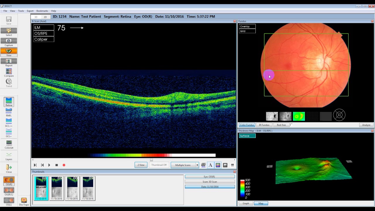
An OCT eye exam is a non-invasive test that provides 3-D color-coded cross sectional images of the retina to enable early detection and treatment of ocular disease that may develop without any noticeable symptoms. B- Scan Display The B-scan display as shown in the above figure right-hand side shows reflection cross-sectional view from the top and bottom of the test object flaws as the probe moves along a line one axis. Line-field confocal optical coherence tomography LC-OCT is an imaging technique based on the principle of time-domain OCT with line illumination using a broadband laser and line detection using a line-scan camera.

Demonstration Of Choroidal Neovascularization Associated With An Intraretinal Lesion On Indocyanine Green Ang Optical Coherence Tomography Eye Health Optometry

Polypoidal Choroidal Vasculopathy Pcv

Figure 1 Vascular Probe Continuity

Pin Page

Ophthalmology Management Cirrus Hd Oct Today And Tomorrow

The Anatomy Of An Oct Scan

Pin Page

Pin On Casa Aperta Daybed

Figure 2 Normal Rnfl Thickness In Optical Coherence Tomography Onh

Macular Pucker Numerous Underlying Conditions Lead To Scar Tissue On The Macula When This Affects Vision The Scarring Can B Scar Tissue Pathology Restoration

Instagram Post By Bhavin Jankharia Oct 22 2015 At 4 27am Utc

Pin On Patient Information Pages
Tyrrells And Embery Opticians Adli Kullanicinin Optical Coherence Tomography Oct Panosundaki Pin

Pin Page

Pin Page

The Official Oct Interpretation

Mesh Stigmergy
Maestro Software Tutorial

Pin On Glaucoma
0 Response to "oct b scan"
Post a Comment